Service
Real Time Ultrasound
See what your muscles are doing in real time with our ultrasound machine. This is one of the best ways to learn how to activate deep muscles and make sure your body is getting the support it needs.

See what your muscles are doing in real time with our ultrasound machine. This is one of the best ways to learn how to activate deep muscles and make sure your body is getting the support it needs.
Learning to activate and perform exercises that focus on muscles close to the surface are difficult enough. When it’s a muscle that is deep down below the surface that you need to learn to use, this can be very challenging indeed. One of the developments in technology that has greatly helped the process of learning how to turn on your deep muscles, is a piece of equipment called a real-time ultrasound.
This ultrasound machine will show you in real time the tightening of your deep muscles so you can know whether you are learning the exercise correctly or not. It is then from this knowledge of how to do the exercise correctly, you can keep practicing this exercise at home.
**As we have specific therapists who are trained to use this real-time ultrasound machine, please contact reception to enquire as to which therapist would be best for you to book with**

Our highly experienced team of physiotherapy practitioners are committed to treating and improving your movement. With the specific training, knowledge and skills to diagnose and treat a wide range of conditions, our team not only focusses on treating your injury but also on helping you to understand how recurrences can be avoided in future.
Learn More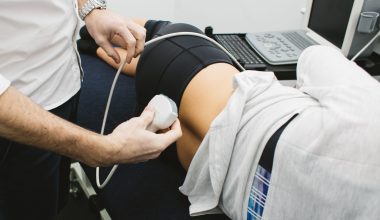
See what your muscles are doing in real time with our ultrasound machine. This is one of the best ways to learn how to activate deep muscles and make sure your body is getting the support it needs.
Learn More
Led by physiotherapists, our Clinical Pilates classes aim to promote movement and strengthening to people of all fitness levels. Whether you're recovering from injury, wanting to learn more about your body, wanting to prevent future injury through strengthening your body or returning to physical activity after time off, we have a Clinical Pilates class or service to help.
Learn More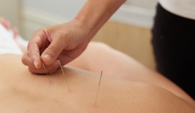
A service offered by all of our physiotherapists and myotherapists, dry-needling involves inserting an acupuncture needle through the skin into a tight muscle to help it relax.
Learn More
Working to stimulate blood flow in the body, Remedial Massage assists in the repair of damaged tissue while also alleviating tightness and tension in the muscles. A practice refined over thousands of years, Remedial Massage is an effective treatment for conditions such as sporting injuries, headaches, muscle cramps and lower back pain.
Learn More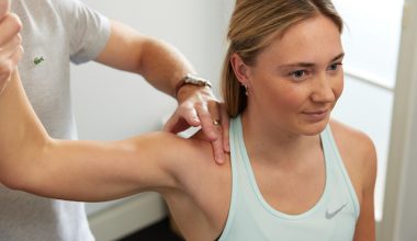
We're proud to offer the services of a Specialist Musculoskeletal Physiotherapist FACP. The highest level of clinical training with only 15 practitioners holding this title in Victoria, Specialist Physiotherapy may be the solution for those suffering from more complex conditions such as persistent low back pain, neck pain, headache and dizziness, and pain conditions such as fibromyalgia.
Learn More
Using one of the latest tools to measure muscle strength and function, this strength assessment helps to ascertain any weaknesses in the body and which exercises will be best to treat them.
Learn More
Get treatment and management for any health concerns such as pelvic floor dysfunction, incontinence, pre- and post-natal care and menopausal changes in the body. Work with our expert practitioner, who holds post-graduate training in pelvic floor physiotherapy.
Learn More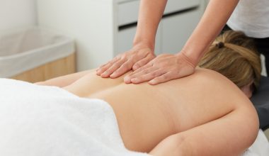
Encompassing treatments such as remedial massage, dry needling, cupping and manual trigger-point therapy, myotherapy offers a holistic, hands-on approach to treating your body. Our Myotherapists' extensive knowledge of anatomy and pathology allows them to tailor a treatment plan specific to your pain or injury.
Learn More
Let us guide you through exercises that are suitable at different stages of pregnancy and motherhood. Targeting conditions such as back pain, pelvic pain and pelvic floor concerns, we'll help you to build and maintain essential muscle strength to navigate the changes to your body both during and after pregnancy.
Learn More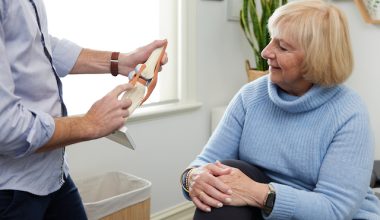
If you have an upcoming surgery, we'll help you to prepare for your best chance of a successful recovery. We take the time to listen to you and educate you about what you'll experience, along with giving you specific exercises to strengthen the relevant muscles ahead of surgery.
Learn More
If you're recovering from major joint surgery, your physio will play a big role in your recovery. Operations such as a knee replacement, hip replacement, shoulder replacement, rotator cuff repair, shoulder stabilisation or knee reconstruction all require expert guidance. Let a physio at Malvern Physio help to plan your treatment at each stage of recovery to help you bounce back as quickly as possible.
Learn More
Get the expert advice and treatment of our practitioners from the comfort of your own home. Conducted via video link, receive the same standard of care as you would in our clinic including a movement assessment and self-management exercise program to treat your concern.
Learn MoreA real-time ultrasound (RTUS) is a diagnostic imaging technique used by physiotherapists to assess and train muscle activation or deactivation. Unlike traditional ultrasound scans used for medical diagnoses, RTUS provides live, moving images of muscles, tendons, and other soft tissues, allowing both the physiotherapist and the patient to see how muscles are working in real-time.
RTUS is particularly useful for assessing deep core muscles, pelvic floor muscles, and other stabilizing muscles that are difficult to feel or activate correctly. It helps with:
By using RTUS, physiotherapists can develop more targeted treatment plans, ensuring patients are correctly engaging the right muscles for recovery and long-term function.
The time it takes to receive ultrasound results in Australia can vary depending on the type of ultrasound, the urgency of the case, and the healthcare provider. However, here are general timeframes:
Routine Ultrasound (e.g., pregnancy, abdominal, musculoskeletal scans): Results are typically available within 24–48 hours. The radiologist interprets the images and sends a report to your referring doctor.
Urgent or Emergency Ultrasound: Results can be available within a few hours, especially if done in a hospital or emergency setting. The radiologist may provide an immediate verbal report to the doctor if urgent action is required.
Specialist or Complex Ultrasounds (e.g., detailed organ scans, Doppler studies): These may take 2–5 days, depending on the complexity and the radiology provider’s workload.
Real-Time Ultrasound for Physiotherapy or Guided Treatment: If performed by a physiotherapist or specialist for assessment, feedback is usually immediate, as they interpret the scan in real time.
Real-Time Ultrasound (RTUS) is a non-invasive imaging technique that uses sound waves to create live images of soft tissues, organs, and muscles. In physiotherapy, RTUS is primarily used to assess muscle function, guide rehabilitation, and monitor treatment progress, rather than for traditional diagnostic purposes like detecting diseases.
However, it is highly effective in identifying musculoskeletal issues, movement dysfunctions, and certain soft tissue abnormalities.
Limitations of Real-Time Ultrasound:
While RTUS is valuable for assessing soft tissues and muscle function, it is not designed to detect:
Real-Time Ultrasound is a valuable tool in physiotherapy for diagnosing muscle dysfunction, soft tissue injuries, and postnatal pelvic health issues. It allows physiotherapists to assess how muscles activate during movement and tailor rehabilitation programs accordingly.
By providing live feedback, RTUS helps patients visualize their muscle engagement and improve their technique during rehab, leading to faster, more effective recovery.
Real-Time Ultrasound (RTUS) is a non-invasive imaging technique that uses sound waves to provide live, dynamic images of muscles, tendons, ligaments, and soft tissues. In physiotherapy, RTUS is used as a biofeedback tool to assess and guide muscle activation, improve movement patterns, and support the rehabilitation process.
While commonly associated with diagnostic imaging in medical fields, in physiotherapy, real-time ultrasound focuses on functional assessment and rehabilitation, rather than diagnosing structural damage.
RTUS allows physiotherapists to visualize how muscles contract in real time.
It helps assess whether key stabilizing muscles (like the deep core muscles or pelvic floor) are being engaged correctly.
Particularly useful for identifying weak or underactive muscles that may not be firing as they should during movement.
Patients can see their muscles working on the screen as they perform movements, providing immediate feedback.
This helps patients learn how to correctly activate specific muscles, such as the transverse abdominis, pelvic floor, or multifidus.
Visual feedback enhances body awareness and helps correct faulty movement patterns that may lead to injury.
RTUS is widely used in the rehabilitation of pelvic floor dysfunction, postnatal recovery, and conditions like urinary incontinence.
It helps assess pelvic floor contractions and guides patients in properly engaging deep core muscles.
In postnatal women, it helps track recovery from diastasis recti (abdominal separation) and guides safe core exercises.
RTUS helps physiotherapists identify muscle imbalances, asymmetries, or compensatory movements that may contribute to pain or dysfunction.
It’s particularly useful in assessing conditions like:
RTUS can visualize tendon injuries, muscle tears, and scar tissue formation to monitor healing progression.
It guides the physiotherapist in adjusting exercise intensity and load as the tissue heals, ensuring a safe return to activity.
By periodically using RTUS throughout rehabilitation, physiotherapists can track improvements in muscle strength, endurance, and control.
This ensures that exercises are effective and allows for real-time adjustments to treatment plans.
Conditions Commonly Supported by Real-Time Ultrasound in Rehabilitation:
Real-Time Ultrasound is a valuable tool in injury rehabilitation, providing both the physiotherapist and the patient with a clear, visual understanding of muscle function and movement patterns. By offering real-time feedback, it empowers patients to engage the right muscles, correct faulty movement patterns, and accelerate recovery.
It is particularly beneficial for conditions involving deep core instability, pelvic floor dysfunction, or chronic pain, where visualizing muscle activation makes a significant difference in rehabilitation outcomes.
If you’re struggling with chronic pain, postnatal recovery, or recurring injuries, ask your physiotherapist whether real-time ultrasound could enhance your rehabilitation process.